1. Disease overview of aspergillosis
Aspergillosis is a fungal infection of Caribbean soft corals. It affects 6 species of sea fan corals and gorgonians widely distributed throughout the Caribbean. The pathogen is an Aspergillus species that infect gorgonians after spore germination on the coral surface. This is followed by the infiltration and spread of the hyphae in the coral tissue, causing visible damage. Biological damage can be associated with complete loss of tissue and bone, often occurring at multiple sites in the infected population. Coral hosts may develop purple galls to encase fungal hyphae. Fungal hyphae are visible if any of these galls are cut open. One known source is African dust.
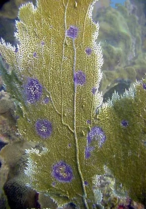
Image: Aspergillosis is caused by the terrestrial Aspergillus salicylate. The disease causes lesions associated with degraded gorgonian tissue. Gorgonian corals fight the disease by encasing fungal hyphae in purple galls.
2. Bacteria-induced bleaching
Bleaching caused by specific bacterial infections (as opposed to environmental stress responses) occurs in the absence of zooxanthellae due to toxins produced by intracellular bacterial pathogens. Bacterial bleaching occurs in Mediterranean reef-building corals. Two known albino pathogens are Vibrio shiloi and V. patogonica. Initial pathogens on corals attach to b-galactose in the polysaccharide layer on the coral host surface. The subsequent temperature-dependent growth of bacteria within the cell leads to an invasion of coral host tissue. The production of a heat-sensitive toxin causes the lysis of the zooxanthellae. Conditions include loss of zooxanthellae with intact coral tissue and, unlike environmental bleaching, the presence of Vibrio shiloi or patogonica in affected tissue. The exact source is unknown.
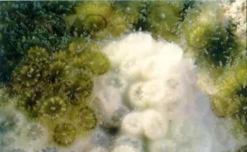
Image: Bacterial bleaching is caused by a specific bacteria/coral interaction. Specificities include pathogen recognition by host (coral) surface receptors; invasion of coral tissue; proliferation of bacteria within coral tissue; release of bacterial toxins that cause coral bleaching.
3. Overview of Black Band Disease
Black band disease is characterized by complete degradation of coral tissue due to a mat of migratory microorganisms that appear dark red or black in the pathogenic microbial community. This mat appears between the surface of healthy coral tissue and newly exposed coral skeleton. Band color may be dark brown to red, depending on the vertical position of the cyanobacterial population associated with the band. The vertical position is based on the light intensity-dependent photoresponse of cyanobacterial filaments, and the color (attributed to the cyanobacterial pigment phycoerythrin) depends on the thickness of the band. The strap is about 1mm thick and has a width from 1mm to 7cm. White spots may appear on the surface, forming dense white patches over time. Pathogenic microbial mats move through coral colonies at rates of 3 mm to 1 cm per day. Tissue death results from exposure to a hypoxic, sulfide-rich microenvironment.
The black belt microbial flora consists of a group of photosynthetic and non-photosynthetic bacteria that coexist synergistically. This flora has three functionally and physically dominant fungi, as well as a number of heterotrophic fungi whose roles in the disease are unknown. The three functionally dominant bacteria were cyanobacteria, sulfate-reducing bacteria and sulfate-oxidizing bacteria.
Black band disease affects 42 species of coral worldwide. The only known source is within cyanobacterial biofilms in sediment depressions of healthy black-band disease-susceptible corals.
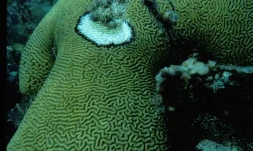
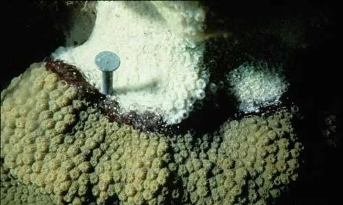
Image: Black band disease is characterized by a black ring, or band, that separates apparently healthy coral tissue from newly exposed coral skeleton. It migrates in coral colonies and completely decomposes coral tissue. A closer look shows that the band is made up of many microorganisms, seen here as a dark photosynthetic cyanobacteria (cyanobacteria) and white-spotted communities of sulfur bacteria.
4. An overview of black spot disease.
Black spot disease appears as dark (brown or purple) pigmented areas of reef-building coral tissue. No known pathogens. Pigmented areas on coral skeletons may or may not be skeletal depressions. Coral tissue remained intact, although damage and death of coral tissue was sometimes observed in the center of the spot. The disease is common in the Caribbean.
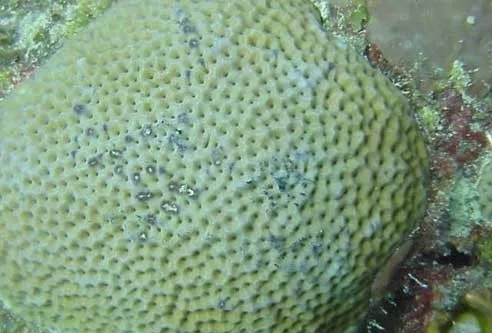
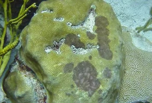
Image: There are no known pathogens of black spot disease, which is identified by dark spots on coral tissue. If present, tissue loss is minimal.
Source:NOAA