5. Overview of leucorrhea:
Leucorrhea is characterized by complete degradation of the coral tissue of Caribbean staghorn corals. There are two species affected, one is the palmophyllum staghorn coral and the other is cervicornis staghorn (Gladfelter, 1982). The disease draws a sharp line between apparently healthy coral tissue and exposed coral skeleton. These symptoms are the same as white blight, except that the white band is unique to staghorn corals (and white blight has not been found in staghorn corals). Tissue loss usually begins from the base of the branch to the tip, but can also start from the middle of the branch. There are two distinct disease types that differ in tissue loss patterns. Leucorrhea type I exhibits tissue degradation associated with lines that migrate throughout coral colonies. Although the newly exposed coral skeletons were banded, there were no distinct microbial bands. Tissue lysis is always associated with a moving leading edge (this is the difference between Type I and Type II. Tissue loss rates vary from mm to cm/day. Type II leucorrhea also exhibits tissue degradation as it travels through coral colonies, but in In this case, movement sometimes occurs in areas of albinism, catching up with active tissue lysis (Ritchie and Smith, 1998). The only way to distinguish the two types is to observe the progression of the bands over time.
Leucorrhea disease affects staghorn corals throughout the Caribbean and causes massive coral die-offs on a regional scale.
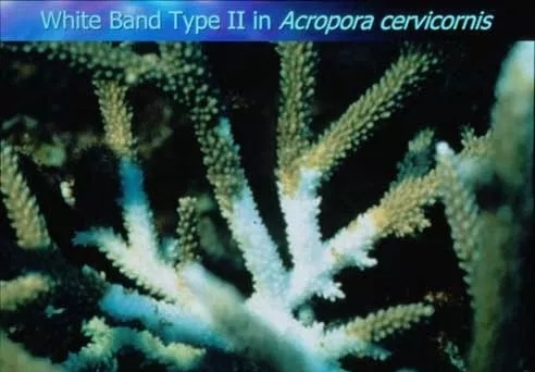
Image: Leucorrhea has two causes, type I and type II. In type I, tissue destruction is associated with anterior movement of the band. In type II, there is sometimes an albino zone between the tissue degradation zone and the shifting peak zone. Type I is visually indistinguishable from type II if there is no whitening zone. No pathogens have been isolated so far.
6. Overview of Leukemia
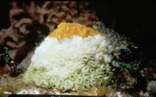
White blight is characterized by a sharp demarcation line between apparently healthy coral tissue and exposed coral skeleton. No obvious microbial zone was seen. The blight is caused by a bacterial pathogen (Aurantimonas coralicida). Disease signs (rates and patterns of disease progression and toxicity) differed between the three different types. Type I blight, recorded in the 1970s and 1980s, started on both sides of the colony, with tissue destruction at a rate of 3 mm per day. Six species of coral are reported to be affected. Type II blight, first recorded in 1995, starts at the bottom of coral colonies and progresses upwards, with tissue damage up to 2 cm/day. Whitened areas (<3 mm) may exist between healthy tissue and exposed bone. Type III blight, which first emerged in 1999, begins at the flanks or top of the colony and destroys tissue at a high rate of dm per day. . White blight is currently endemic throughout the Caribbean, affecting 33 species of Caribbean stony corals (Weil et al, in press). The source is not yet known.
7. Overview of white pox
White pox is characterized by the degradation of coral tissue, the rapid loss of coral tissue along a distinct line, or the presence of small residual tissue anywhere above or below branches, sometimes near the edges of irregularly shaped patches. The average tissue loss rate is 2.5 cm2/day, but can be as high as 10.5 cm2/day.
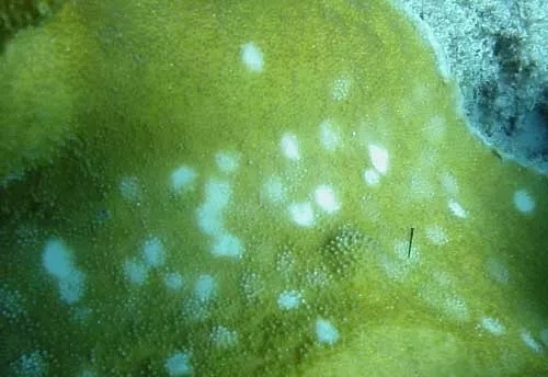
Image: White pox is characterized by round lesions. The pathogen is Serratia marcescens, a gram-negative enterobacter
8. Overview of Yellow Belt Disease
Yellow band disease is characterized by large rings or patches of bleached yellow tissue in Caribbean reef-building corals. It spreads widely in the Caribbean. Tissue loss is extremely slow (cm/year). When yellow-banded corals also suffer from bleaching, the yellow-banding can be mixed with bleached features; after recovery from bleaching, the yellow-banding is visible again. Although loss of zooxanthellae pigments and zooxanthellae cells has been found in affected tissues, no pathogen has been identified. Zooxanthellae associated with the yellow belt have a lower mitotic index (the number of dividing cells), and it has been suggested that the disease affects zooxanthellae rather than corals.
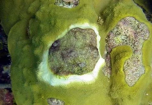
Image: The yellow band is characterized by a light-like expansion of the albino band. Minimal tissue loss (cm/year). No pathogens were isolated.
Source: NOAA